human muscular structure, computer artwork. - cell membrane stock illustrationsHuman muscular structure, computer artwork.
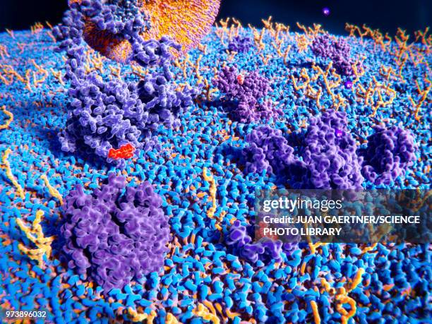
transmembrane protein, illustration - cell membrane stock illustrationsTransmembrane protein, illustration
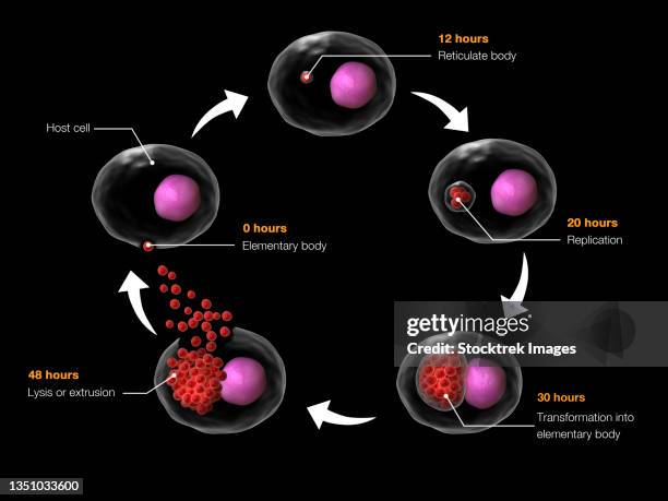
life cycle of chlamydia psittaci. - cell membrane stock illustrationsLife cycle of Chlamydia psittaci.
nicotinic acetylcholine receptor, illustration - cell membrane stock illustrationsNicotinic acetylcholine receptor, illustration
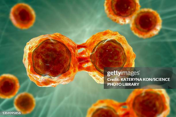
cancer cells dividing, conceptual illustration - cell membrane stock illustrationsCancer cells dividing, conceptual illustration
conceptual image of radiolarians with a skeletal frame. radiolarians are tiny protozoans that live in the ocean. - cell membrane stock illustrationsConceptual image of Radiolarians with a skeletal frame. Radiolarians are tiny protozoans that live in the ocean.
bacterial inclusion bodies, illustration - cell membrane stock illustrationsBacterial inclusion bodies, illustrationFlow Chart
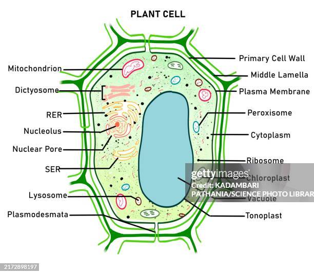
plant cell, illustration - cell membrane stock illustrationsPlant cell, illustrationCytoplasm, Illustration, Plant Cell
l,d-transpeptidase bound to peptidoglycan, illustration - cell membrane stock illustrationsL,D-transpeptidase bound to peptidoglycan, illustration
cell membrane structure, illustration - cell membrane stock illustrationsCell membrane structure, illustration
covid-19 coronavirus entering the lungs. - cell membrane stock illustrationsCOVID-19 coronavirus entering the lungs.
illustration of action potential of a nerve cell. - cell membrane stock illustrationsIllustration of action potential of a nerve cell.Motion
b cell receptor dimer, illustration - cell membrane stock illustrationsB cell receptor dimer, illustration
palmitoyloleoyl pe lipid bilayer - cell membrane stock illustrationsPalmitoyloleoyl PE lipid bilayerPope, Illustration
zosurabalpin antibiotic action, illustration - cell membrane stock illustrationsZosurabalpin antibiotic action, illustration
pten-induced kinase 1 bound to mitochondrion, illustration - cell membrane stock illustrationsPTEN-induced kinase 1 bound to mitochondrion, illustration
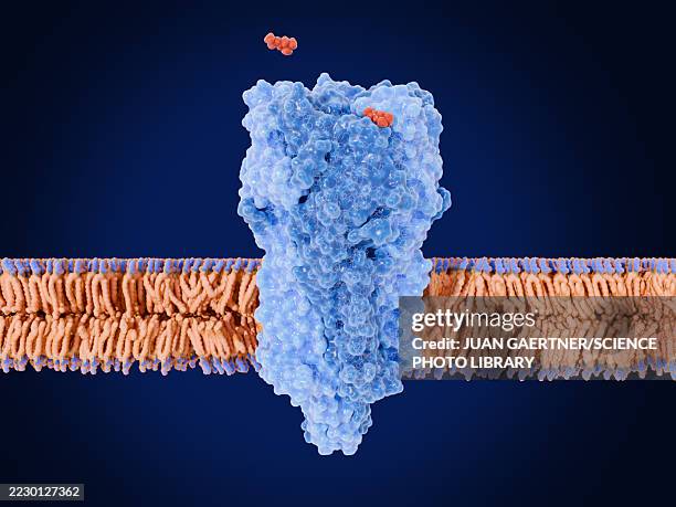
oxycodone and opioid receptor, illustration - cell membrane stock illustrationsOxycodone and opioid receptor, illustration
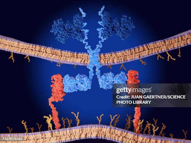
her2 and her3 cell membrane receptors forming complex, illustration - cell membrane stock illustrationsHER2 and HER3 cell membrane receptors forming complex, illustrationProtein
structure of a generic centric diatom, illustration - cell membrane stock illustrationsStructure of a generic centric diatom, illustration
coxsackievirus bound to human cell, illustration - cell membrane stock illustrationsCoxsackievirus bound to human cell, illustrationInfectious Disease, Human Cell
vancomycin resistance protein, molecular model - cell membrane stock illustrationsVancomycin resistance protein, molecular model
amitriptyline antidepressant drug action, illustration - cell membrane stock illustrationsAmitriptyline antidepressant drug action, illustration
chimeric antigen receptor (car) therapy, illustration - cell membrane stock illustrationsChimeric antigen receptor (CAR) therapy, illustration
styrene maleic acid lipid particle, illustration - cell membrane stock illustrationsStyrene maleic acid lipid particle, illustration
animal cell, illustration - cell membrane stock illustrationsAnimal cell, illustrationMitochondrion, Illustration, Biology
car t cell cancer immunotherapy, illustration - cell membrane stock illustrationsCAR T cell cancer immunotherapy, illustrationT-Cell, Antigen, Immunotherapy
t cell receptor, cd4 and mhc proteins, illustration - cell membrane stock illustrationsT cell receptor, CD4 and MHC proteins, illustrationProtein, The Human Body, Three Dimensional
stem cells, illustration - cell membrane stock illustrationsStem cells, illustrationBiomedical Illustration
amyloid precursor protein at cell membrane, illustration - cell membrane stock illustrationsAmyloid precursor protein at cell membrane, illustrationProtein, Brain, Molecule
car t cell cancer immunotherapy, illustration - cell membrane stock illustrationsCAR T cell cancer immunotherapy, illustrationScience, T-Cell, Immunology
glp-1 receptor activated by semaglutide, illustration - cell membrane stock illustrationsGLP-1 receptor activated by semaglutide, illustrationObesity, Three Dimensional, Semaglutide
ldl receptors on cell membrane, illustration - cell membrane stock illustrationsLDL receptors on cell membrane, illustrationLipoprotein, Triglyceride, High Density Lipoprotein
rage receptors in membrane, illustration - cell membrane stock illustrationsRAGE receptors in membrane, illustrationProtein, Diabetes, Molecule
stem cells, illustration - cell membrane stock illustrationsStem cells, illustrationLymphoma, Biology, Human Cell
microscopic view of the covid-19 coronavirus. - cell membrane stock illustrationsMicroscopic view of the COVID-19 coronavirus.
human muscular structure, computer artwork. - cell membrane stock illustrationsHuman muscular structure, computer artwork.Muscle Fibre
zosurabalpin antibiotic action, illustration - cell membrane stock illustrationsZosurabalpin antibiotic action, illustration
activation of t-cell immune response, illustration - cell membrane stock illustrationsActivation of T-cell immune response, illustration
interleukin 4 binding to its receptor, illustration - cell membrane stock illustrationsInterleukin 4 binding to its receptor, illustration
upper cell membrane, illustration - cell membrane stock illustrationsUpper cell membrane, illustration
gram negative bacterium, illustration - cell membrane stock illustrationsGram negative bacterium, illustration
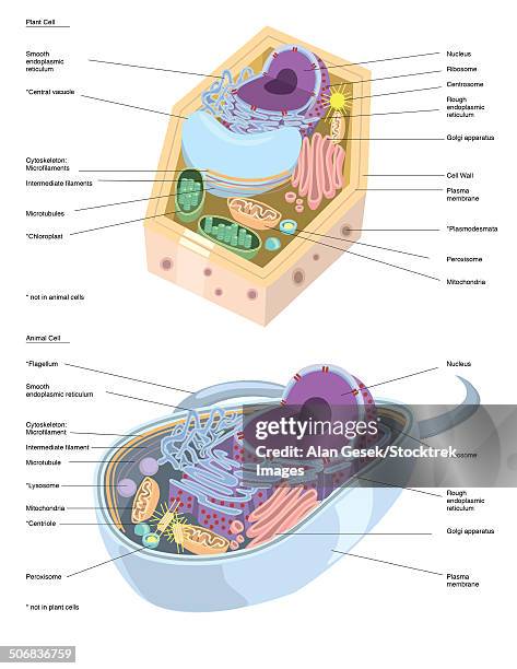
comparative illustration of plant and animal cell anatomy (with labels). - cell membrane stock illustrationsComparative illustration of plant and animal cell anatomy (with labels).
rod-shaped gram-negative bacterium, illustration - cell membrane stock illustrationsRod-shaped Gram-negative bacterium, illustrationE. Coli
cross section of the covid-19 coronavirus. - cell membrane stock illustrationsCross section of the COVID-19 coronavirus.
cell membrane lipid bilayer, artwork - cell membrane stock illustrationsCell membrane lipid bilayer, artwork
conceptual illustration of rh factor on a red blood cell. - cell membrane stock illustrationsConceptual illustration of Rh factor on a red blood cell.
structure of animal cell, illustration - cell membrane stock illustrationsStructure of animal cell, illustrationCell Organelle, Cytoplasm
escherichia coli anatomy, illustration - cell membrane stock illustrationsEscherichia coli anatomy, illustrationE. Coli
somatostatin binding to somatostatin receptor, illustration - cell membrane stock illustrationsSomatostatin binding to somatostatin receptor, illustration