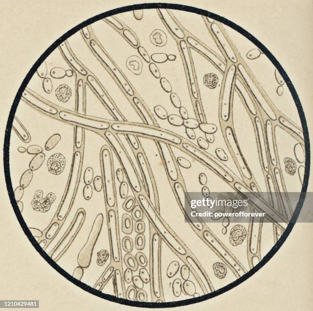
microscopic view of candida albicans from a patient with mycosis - 19th century - yeast microscope stock illustrationsMicroscopic View of Candida Albicans from a Patient with Mycosis - 19th CenturyArchival, Engraved Image, Illustration
a scientific diagram showing microscopic yeast cells budding and forming chains within a circular frame - yeast microscope stock illustrationsA scientific diagram showing microscopic yeast cells budding and forming chains within a circular frame
yeast trpy1 channel with lipid and calcium, illustration - yeast microscope stock illustrationsYeast TRPY1 channel with lipid and calcium, illustration
yeast pheromone receptor dimer bound to two g proteins, illustration - yeast microscope stock illustrationsYeast pheromone receptor dimer bound to two G proteins, illustration
skin with cutaneous candidiasis, illustration - yeast microscope stock illustrationsSkin with cutaneous candidiasis, illustration
a technical diagram displaying microscopic view of bottom yeast cells with granulated textures inside a circular border - yeast microscope stock illustrationsA technical diagram displaying microscopic view of bottom yeast cells with granulated textures inside a circular border
yeast trpy1 channel with lipid and calcium, illustration - yeast microscope stock illustrationsYeast TRPY1 channel with lipid and calcium, illustration