cross section illustration of human large intestine - serosa stock illustrationsCross section illustration of human large intestine
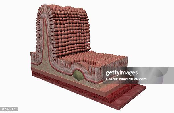
cross section illustration of human small intestine showing muscle layer, villus and plicae - serosa stock illustrationsCross section illustration of human small intestine showing muscle layer, villus and plicae
small intestine and attached mesentery, 10x - serosa stock pictures, royalty-free photos & imagesSmall Intestine and attached Mesentery, 10xSmall Intestine, Smooth Muscle, Immunofluorescent Photomicrograph
cross-section of stomach, illustration - serosa stock illustrationsCross-section of stomach, illustration
cross-section of stomach, illustration - serosa stock illustrationsCross-section of stomach, illustration
uterus after endometrium sloughed off, illustration - serosa stock illustrationsUterus after endometrium sloughed off, illustration
endometriosis blocking egg in fallopian tube, illustration - serosa stock illustrationsEndometriosis blocking egg in fallopian tube, illustrationEndometriosis
backflow of endometrial lining, illustration - serosa stock illustrationsBackflow of endometrial lining, illustrationEndometriosis, Medicine, Healthcare And Medicine
ovum moving through ampulla and isthmus, illustration - serosa stock illustrationsOvum moving through ampulla and isthmus, illustration
ovum travelling into uterine cavity, illustration - serosa stock illustrationsOvum travelling into uterine cavity, illustration
ovum travelling through fallopian tube, illustration - serosa stock illustrationsOvum travelling through fallopian tube, illustration
ovum inside uterine cavity, illustration - serosa stock illustrationsOvum inside uterine cavity, illustration
ovum exiting through vagina, illustration - serosa stock illustrationsOvum exiting through vagina, illustration
layers of small intestine, illustration - serosa stock illustrationsLayers of small intestine, illustration
old engraved illustration of a serous membrane (or serosa) a thin, two-layered membrane lining the body cavities and covering the organs within them - serosa stock pictures, royalty-free photos & imagesOld engraved illustration of a serous membrane (or serosa) a thin, two-layered membrane lining the body cavities and covering the organs within them
old engraved illustration of a serous membrane (or serosa) a thin, two-layered membrane lining the body cavities and covering the organs within them - serosa stock pictures, royalty-free photos & imagesOld engraved illustration of a serous membrane (or serosa) a thin, two-layered membrane lining the body cavities and covering the organs within themDrawing - Artistic Product, Engraved Image
old engraved illustration of a serous membrane (or serosa) a thin, two-layered membrane lining the body cavities and covering the organs within them - serosa stock pictures, royalty-free photos & imagesOld engraved illustration of a serous membrane (or serosa) a thin, two-layered membrane lining the body cavities and covering the organs within them
stomach with cancer tissue - serosa stock illustrationsStomach With Cancer TissueStomach Cancer, Gastric Ulcer, Stomach
anatomy of the structure and layers of the stomach wall. - serosa stock illustrationsAnatomy of the structure and layers of the stomach wall.
anatomy of the structure and layers of the stomach wall. - serosa stock illustrationsAnatomy of the structure and layers of the stomach wall.Mucus
Digestive System, Illustration, The Digestive Tube, Extending From The Esophagus To The Anal Canal, Is Composed Of A Wall Containing Four Tunics, Or...Digestive System, IllustrationMesentery
mesothelium, visceral peritoneum and serosal layer - serosa stock pictures, royalty-free photos & imagesMesothelium, Visceral Peritoneum and Serosal layer
peyer's patch--aggregated lymphatic nodules, ileum - serosa stock pictures, royalty-free photos & imagesPeyer's Patch--aggregated Lymphatic Nodules, Ileum
colon--mucosa, submucosa, muscularis, serosa, 25x - serosa stock pictures, royalty-free photos & imagesColon--Mucosa, Submucosa, Muscularis, Serosa, 25X
Model Of The Internal Anatomy Of An Adult Female Genitourinary Tract Anterior View Of A Median Section. The Urinary Bladder And The Uterus Lie In...Anatomy, Female Genitalia
Model Of The Internal Anatomy Of An Adult Human Male Genitourinary Tract Anterior View Of A Median Section. The Urinary Bladder And The Prostate Lie...Anatomy, Male Genitalia
simple squamous epithelium, small intestine, 250x - serosa stock pictures, royalty-free photos & imagesSimple Squamous Epithelium, Small Intestine, 250X
small intestine layers--mucosa, submucosa, muscularis, serosa--jijunum 50x - serosa stock pictures, royalty-free photos & imagesSmall Intestine layers--Mucosa, Submucosa, Muscularis, Serosa--Jijunum 50X
Diagram Of The Human Large Intestines, Including And Inset Of Its Musculature And Mucosa Histology And An Inset Of Its Regions.Diagram Of The Human Large Intestines, Including And Inset Of Its Musculature And Mucosa Histology And An Inset Of Its Regions
Diagram Of The Human Small Intestine, With Insets Of Musculature, Mucosa Histology And Its Regions In Situ.Diagram Of The Human Small Intestine, With Insets Of Musculature, Mucosa Histology And Its Regions In SituSmall
Diagram Showing The Mucosa And Musculature Of The Human Stomach Plus Insets Of Histology And Regions.Diagram Showing The Mucosa And Musculature Of The Human Stomach Plus Insets Of Histology And RegionsStomach
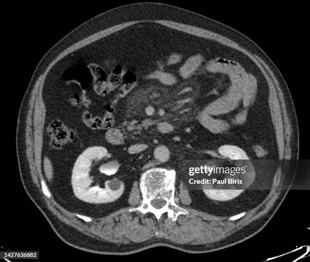
ct findings in sclerosing mesenteritis (panniculitis), coronal view - serosa stock pictures, royalty-free photos & imagesCT Findings in Sclerosing Mesenteritis (Panniculitis), coronal view
ct findings in sclerosing mesenteritis (panniculitis), axial view - serosa stock pictures, royalty-free photos & imagesCT Findings in Sclerosing Mesenteritis (Panniculitis), axial viewMedical X-ray, Abdomen, Colon
large intestine (colon), layers (or tunics), 10x. shows: mucosa, submucosa, muscularis (smooth muscle - longitudinal and circular layers), serosa, goblet cells (red). - serosa stock pictures, royalty-free photos & imagesLARGE INTESTINE (COLON), LAYERS (OR TUNICS), 10X. Shows: mucosa, submucosa, muscularis (smooth muscle - longitudinal and circular layers), serosa, goblet cells (red).
small intestine (ileum) with villi and layers (or tunics), 25x shows: mucosa, submucosa, muscularis (circular and longitudinal layers), serosa, intestinal glands, and goblet cells. cross section. - serosa stock pictures, royalty-free photos & imagesSMALL INTESTINE (ILEUM) WITH VILLI AND LAYERS (OR TUNICS), 25X Shows: mucosa, submucosa, muscularis (circular and longitudinal layers), serosa, intestinal glands, and goblet cells. Cross section.
small intestine with villi and layers (or tunics) 25x. shows: villi, simple columnar epithelium, goblet cells with mucous, submucosa, muscularis (smooth muscle--circular and longitudinal layers) serosa. - serosa stock pictures, royalty-free photos & imagesSMALL INTESTINE WITH VILLI AND LAYERS (OR TUNICS) 25X. Shows: villi, simple columnar epithelium, goblet cells with mucous, submucosa, muscularis (smooth muscle--circular and longitudinal layers) serosa.
smooth muscle--longitudinal and circular layers - serosa stock pictures, royalty-free photos & imagesSmooth Muscle--Longitudinal and Circular LayersMacrophotography
villi.ileum.shows simple columnar epithelium,goblet cells, striated border,submucosa,serosa,25x muscularis - serosa stock pictures, royalty-free photos & imagesVILLI.ILEUM.SHOWS SIMPLE COLUMNAR EPITHELIUM,GOBLET CELLS, STRIATED BORDER,SUBMUCOSA,SEROSA,25X MUSCULARIS